

For myoblast culture teams, I would select a serum-free medium by two outcomes: viable cell expansion and retained myotube-forming capacity. Start with your cell line’s history and measurable acceptance limits - not a universal recipe.
I would build the workflow around these checks:
- Define permitted inputs. Serum-free does not mean chemically defined or animal-component-free; check supplements and coatings or dissociation reagents.
- Compare basal media and supplements under matched conditions. Track nutrient use, pH and osmolality alongside growth, myogenic markers and premature fusion.
- Test medium and handling together. For adherent cells, qualify the coating–medium pair, seeding density, passaging and recovery. Validate suspension conditions separately.
- Confirm performance across passages and lots. Measure doubling time, viable yield, contamination and differentiation under a fixed induction protocol.
- Keep sourcing records. Use Cellbase to compare supplier specifications, then qualify components yourself; check production-scale supply and food-grade suitability where required.
My selection rule is simple: retain the simplest tested formulation that meets your line-specific limits. <u>More cells alone is not enough.</u>
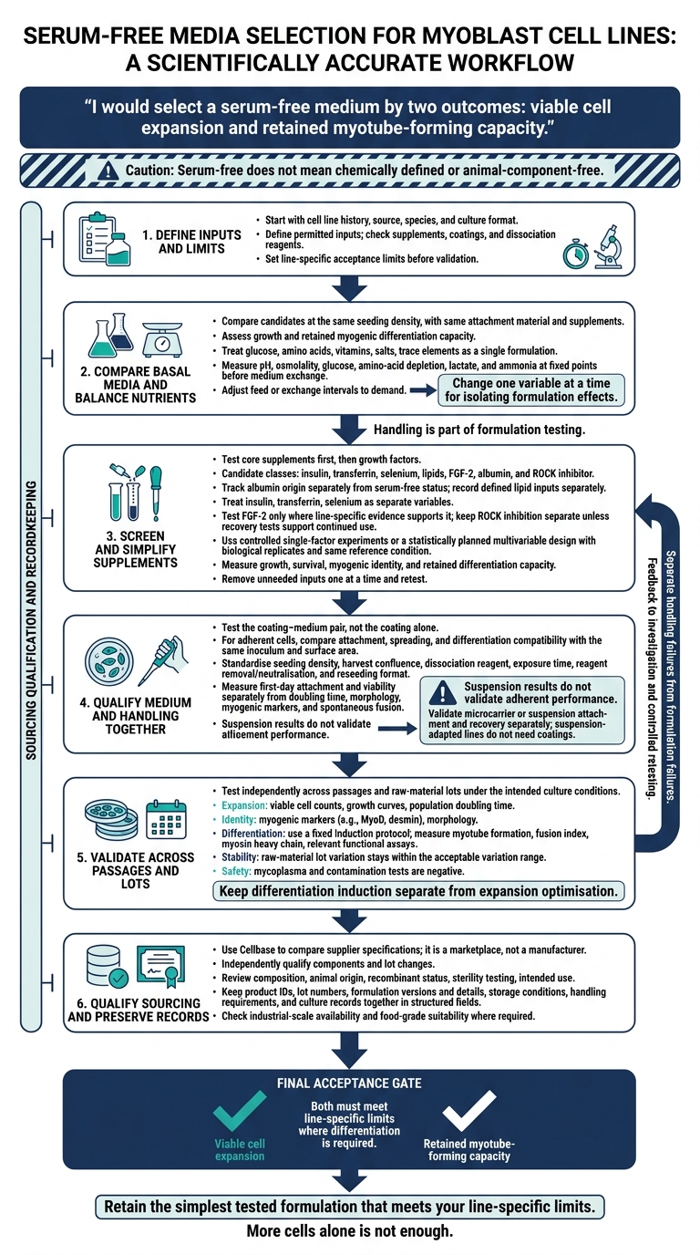
Serum-Free Myoblast Media Design Workflow
Choose the Basal Medium and Nutrients
Shortlist basal media by myoblast source, species and culture format - not by medium name alone. Use the expansion target to rank candidates, and select a medium that supports growth while retaining myogenic potential. Suspension results do not validate adherent performance.[7]
Compare Basal Media
Compare candidates at the same seeding density, with the same attachment material and supplements.[2] Prioritise workflows that demonstrate serum-free expansion in the same cell line while preserving differentiation capacity.
| What to compare | What to check |
|---|---|
| Cell source and species match | Whether the same cell line was used |
| Culture format | Whether the workflow used adherent or suspension-adapted conditions |
| Serum-free expansion | Growth under defined conditions in the relevant cell line |
| Post-expansion phenotype | Retention of myogenic differentiation capacity |
Keep adherent screening results separate from suspension-process validation. Use the best-performing basal medium as the baseline for nutrient balancing.
Balance Nutrients, pH and Osmolality
Treat glucose, amino acids, vitamins, salts and trace elements as a single formulation. Change one variable at a time to isolate its effects on growth and phenotype.
During high-density culture, measure pH, osmolality, glucose, amino-acid depletion, lactate and ammonia. Sample at fixed points before medium exchange, then adjust feed or exchange intervals to match demand.
Once the baseline is stable, screen defined supplements against this formulation.
sbb-itb-ffee270
Choose and Source Defined Supplements
Once the basal medium is fixed, test defined supplements.
Choose Carriers, Lipids and Growth Factors
Track albumin origin separately from serum-free status. Check its composition and record defined lipid inputs separately so you can distinguish their contribution. Treat insulin, transferrin and selenium as separate variables, even when supplied together.
Screen each supplement for improved growth while checking that cells retain their myogenic phenotype.
| Supplement class | Purpose | Expected benefit | Potential drawback | Confirming assay |
|---|---|---|---|---|
| Insulin | Glucose uptake and signalling | Increased proliferation | Requires recombinant sourcing | Proliferation assay |
| Transferrin | Iron delivery | Improved cell survival | Risk of animal origin if not defined | Proliferation rate |
| Selenium | Antioxidant support | Reduced oxidative stress | Toxicity at high concentrations | ROS assay |
| Lipids | Membrane synthesis | Improved growth/phenotype | Poor solubility/stability | Lipid droplet staining |
| FGF-2 | Mitogenic signalling | Fast expansion | Can inhibit differentiation | Myogenic marker expression (e.g. MyoD) |
| Albumin | Carrier for lipids/factors | Improved stability/transport | Batch variability | Growth and viability assay |
| ROCK inhibitor | Recovery aid | Improved post-thaw survival | Potential off-target effects | Colony forming unit (CFU) assay |
Test FGF-2 only where line-specific evidence supports it. Species-specific responses make results uncertain when transferred between lines.[1] Keep ROCK inhibition separate from the routine expansion formulation unless recovery tests support continued use.
Test Combinations and Remove Unneeded Inputs
Test core supplements in the basal medium first, then growth factors. Use controlled single-factor experiments to identify individual effects, or statistically planned multivariable design to assess interactions. Include biological replicates and the same reference condition in every run.
Measure more than metabolic activity. Assess growth and survival alongside myogenic identity and retained differentiation capacity.
Remove unneeded inputs one at a time, then retest each simplified formulation. Retain a component only when retesting confirms that it improves expansion without weakening myogenic identity. For commercial production, also check food-grade suitability and industrial-scale availability; small-scale growth performance alone is not enough.[6]
Source and Qualify Components Through Cellbase
Use Cellbase to source cultivated meat media, supplements and attachment materials. It is a marketplace, not a manufacturer. Its structured metadata supports like-for-like comparisons of specifications.[2]
Review supplier documents for composition, animal origin, recombinant status, sterility testing and intended use. For each version, record product IDs, lot numbers, formulation details, storage conditions and handling requirements.
Treat each listing as a starting point, not proof of suitability. Independently qualify components and lot changes against your acceptance criteria before routine use.
Standardise Attachment and Passaging
Once the formulation is fixed, standardise attachment and passaging so handling differences do not mask medium effects. Both are part of the formulation test, not separate steps.
Test Attachment Materials with the Medium
Test the coating–medium pair, not the coating alone. For adherent myoblasts, compare attachment, spreading and compatibility with differentiation using the same inoculum and surface area. Record the coating’s origin and lot details: serum-free medium does not make an animal-derived coating animal-free.
| Attachment material | Advantage | Limitation to check |
|---|---|---|
| Collagen | Supports attachment and spreading | Often animal-derived; qualify batch consistency |
| Laminin | Supports myoblast attachment | Source and composition can vary |
| Fibronectin | Supports adhesion and spreading | Confirm compatibility with the medium and differentiation workflow |
| Defined extracellular-matrix coatings | Specified composition makes comparisons clearer | Defined does not always mean animal-free |
| Recombinant adhesion proteins | Can reduce reliance on animal-derived materials | Verify line-specific performance and media compatibility |
For microcarriers or suspension, validate attachment and recovery separately; coating data do not transfer. Suspension-adapted lines do not need coatings.[7]
Set Passaging and Recovery Targets
After supplement screening, fix the surface and harvest workflow before comparing growth rates. Using a serum-free media optimization kit can help identify the most effective growth factors during this phase.
Set the starting seeding density and harvest confluence using the line’s validated workflow. Standardise the dissociation reagent, exposure time, neutralisation or removal step, and reseeding format. Use a defined dissociation reagent and verify recovery.
Set a harvest threshold that avoids prolonged over-confluence. Measure first-day attachment and viability, followed by doubling time, morphology, myogenic markers and spontaneous fusion. Passage number alone does not describe culture health. Keep these measurements separate: poor attachment can reduce recovered yield without showing that the medium slows proliferation. Handling and disaggregation methods can also affect yield.[5]
Troubleshoot Growth and Phenotype Changes
Compare candidate and reference media under identical handling conditions, then change one suspected cause at a time. Elongation alone does not establish differentiation; assess myogenic markers and fusion alongside morphology.
When results change, separate handling failures from formulation failures before changing supplements.
| Observation | Possible formulation or handling cause | Discriminating measurement | Corrective experiment |
|---|---|---|---|
| Poor attachment | Coating–medium mismatch; cell damage during handling | First-day attached fraction and viability | Compare coatings using the same medium and inoculum |
| Slow growth | Poor serum-free adaptation; unsuitable density | Doubling time after attachment, at matched density | Compare candidate and reference media using identical handling |
| Death after passaging | Excessive dissociation exposure; residual reagent | Viability immediately after dissociation and the next day | Shorten exposure; validate reagent removal |
| Phenotype drift | Over-confluence; density effects; coating or medium lot change | Confluence, fusion index, myogenic markers, matched-density images and lot records | Passage earlier; retest the previous qualified lot under matched conditions |
| Reduced differentiation capacity | Expansion conditions weakening myogenic capacity | Marker expression and fusion after the same differentiation challenge | Compare expansion formulations at matched density and passage history |
Use the same handling conditions during validation.
Validate the Medium Against Acceptance Criteria
Measure Expansion and Differentiation
Once the workflow is fixed, validate the finished medium under the intended culture conditions. Track viable cell counts and growth curves to measure population doubling time across passages. Assess morphology, myogenic markers and contamination, including mycoplasma, under the intended expansion conditions. Use these results to set the pass/fail limits below.
Keep differentiation induction separate from expansion optimisation. Apply a fixed induction protocol and measure myotube formation, fusion index, myosin heavy chain and relevant functional assays. Retain formulations that preserve differentiation capacity where the workflow requires it.
Define Acceptance Criteria and Transfer Records
Set line-specific limits before validation. Then test them in independent experiments across passages and raw-material lots. Performance under development conditions does not establish transferability, especially when navigating cultivated meat scaling challenges.[4]
| Test | Measurement | Acceptance criterion | Action on failure |
|---|---|---|---|
| Expansion | Viable cell count and doubling time | Meets the line-specific expansion target | Retest mitogenic factors |
| Identity | Myogenic markers such as MyoD and desmin, plus morphology | Expected marker profile and morphology are maintained | Investigate drift or contamination |
| Differentiation | Myotube formation, fusion index, myosin heavy chain and functional assays | Differentiation meets the defined limits under fixed induction conditions | Retest the induction medium |
| Stability | Raw-material lot variation | Performance stays within the acceptable variation range | Qualify a new lot |
| Safety | Mycoplasma and contamination tests | Negative results | Sterilise equipment and discard the batch |
Keep formulation versions, raw-material lots, storage conditions and culture records together in structured fields.[2]
Conclusion: Use the Simplest Validated Formulation
Use the simplest formulation that meets the line-specific criteria, and record it in Cellbase-supported sourcing records.[2]
FAQs
How do I adapt myoblasts to serum-free media?
Move myoblast cultures to defined, animal-free media that support proliferation and differentiation. Screen amino acids, vitamins, inorganic salts, carbohydrates, and recombinant growth factors or cytokines to replace the functions of foetal bovine serum [1][2]. Some researchers use genetic modifications, such as NF2 knockout, to increase proliferation and support adaptation from adherent to suspension culture [3].
Source media components, growth factors and specialised cell lines through Cellbase, a dedicated B2B marketplace for cultivated meat [4].
How many passages should I test before validating a medium?
For a new serum-free medium for myoblast expansion, testing across at least three to five passages is generally recommended, although requirements vary by cell line. Check that proliferation, morphology and differentiation control remain consistent, without relying on residual components carried over from previous media.
Basal media, growth factors and attachment aids for these trials are available through Cellbase, a curated marketplace for the cultivated meat industry.
How can I detect early loss of myogenic potential?
During expansion, regularly monitor proliferation rates, cell morphology and differentiation capacity. Changes in these indicators may suggest that cells are losing their ability to form muscle tissue [1].
Myoblast lines respond to their culture conditions, so match media inputs to your cell line’s requirements [2]. Cellbase offers verified media components, growth factors and analytical tools to help maintain stable culture conditions and support long-term myogenic potential [3].


