

When designing scaffolds for cultivated meat, surface topography is critical for guiding cell growth, alignment, and differentiation. Micro-scale features (1 μm to hundreds of μm) and nano-scale features (10–100 nm) each play distinct roles in shaping cellular behaviour. Micro-topographies influence physical alignment and cell organisation, while nano-topographies work at a molecular level, affecting protein interactions and differentiation pathways.
Key Takeaways:
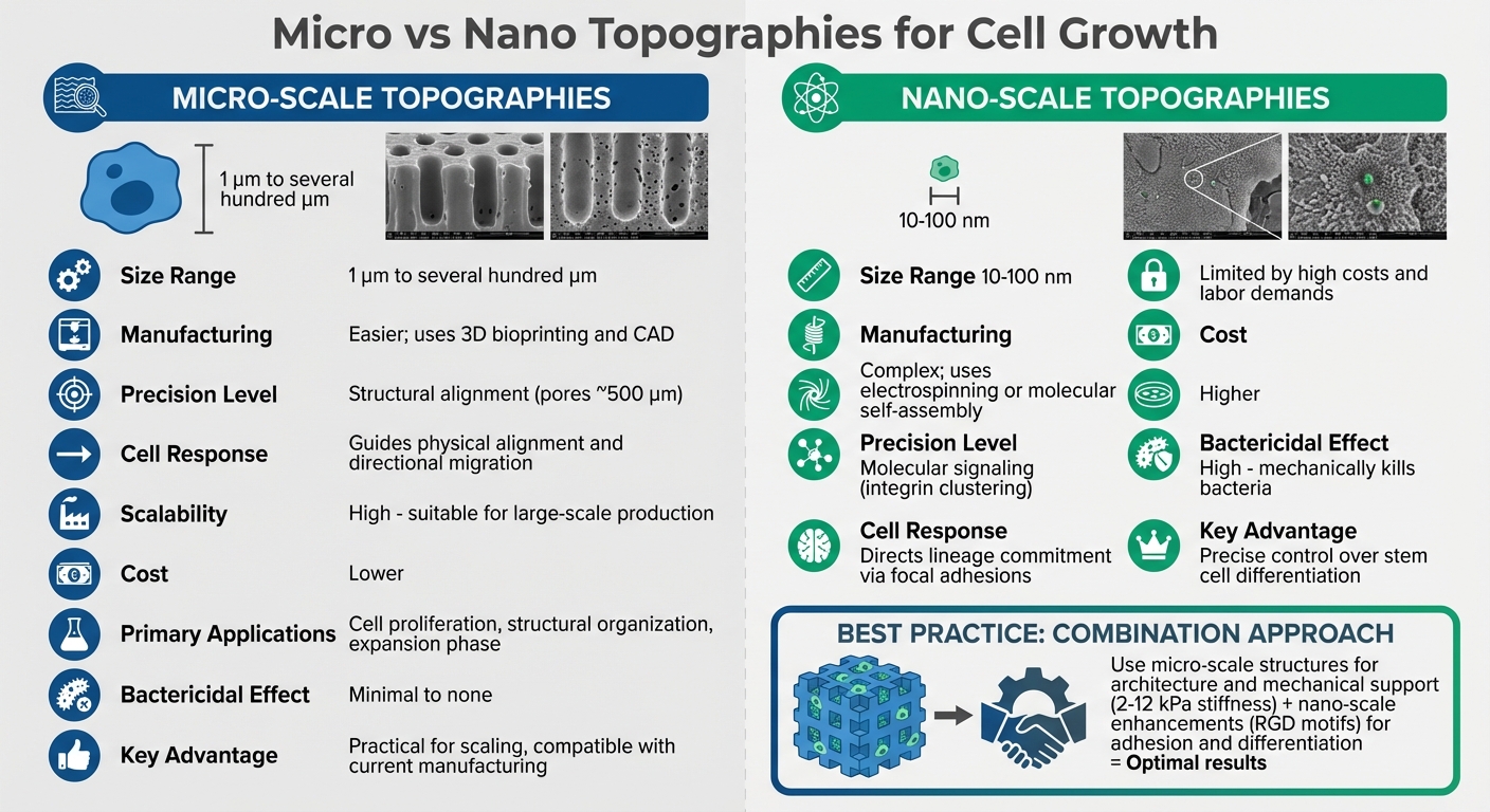
- Micro-scale features: Easier to produce, cost-effective, and suitable for large-scale production. Ideal for cell proliferation and structural organisation.
- Nano-scale features: Mimic natural extracellular matrices, enhancing cell signalling and differentiation but are costlier and harder to scale.
- Combination approach: Using micro-scale structures for architecture and nano-scale enhancements for adhesion and differentiation yields the best results.
Quick Comparison:
| Factor | Micro-Scale Topographies | Nano-Scale Topographies |
|---|---|---|
| Size | 1 μm to several hundred μm | 10–100 nm |
| Manufacturing | Easier, uses 3D bioprinting | Complex, uses electrospinning |
| Precision | Structural alignment | Molecular signalling |
| Scalability | High | Limited |
| Cost | Lower | Higher |
| Applications | Proliferation, alignment | Differentiation, adhesion |
Both approaches have strengths and limitations. Micro-topographies are practical for scalability, while nano-topographies offer advanced control over cellular processes. The best scaffolds often combine these features to optimise cell growth and tissue quality.
Micro vs Nano Scale Topographies for Cultivated Meat Scaffolds Comparison
1. Micro-Scale Topographies
Definition and Characteristics
Micro-scale topographies refer to surface features that range from 1 μm to several hundred micrometres, making them comparable in size to individual cells or larger [3]. These features include structures like micropillars, micro-grooves, and micro-pits, which serve as physical cues that cells interpret through mechanosensing.
One critical factor in how cells respond to these features is surface curvature. For example, micropillars with higher curvature can feel "stiffer" to cells, even if the material itself hasn't changed. This is due to the way non-coplanar forces interact with the cells, creating the perception of increased stiffness [3]. These physical cues have a direct impact on cell shape, growth patterns, and how tissues organise themselves.
Effects on Cell Morphology
Micro-scale features play a significant role in shaping and aligning cells. For instance, fibroblast migration is influenced by pillar spacing between 5 and 10 μm, as this spacing reorganises the actin cytoskeleton. Similarly, increasing the height of micropillars from 1 to 10 μm can enhance laminin expression, which, in turn, affects fibroblast adhesion and morphology [3]. HeLa cells, which are about 4 μm thick, tend to interact primarily with the lower portions of taller pillars, such as those measuring 15.4 μm in height [3].
Effects on Proliferation and Differentiation
The geometry of micropillars also affects cell cycle progression. For example, experiments with PDMS substrates showed that micropillars with a height of 15.4 μm and base diameters between 17.4 μm and 43.9 μm altered the proportion of cells in the S-phase [3]. This ability to control proliferation rates is particularly important for scaling up cultivated meat production.
Micro-scale confinement can also mimic natural tissue organisation. For example, confined microenvironments encourage lumen formation in epithelial and endothelial cells [5], guiding cells to form tissue-like structures. While cells on flat surfaces tend to form monolayers, specific confinement patterns can lead to more complex, three-dimensional arrangements. This control over cell behaviour is crucial for designing scaffolds that support the development of cultivated meat.
Implications for Cultivated Meat Scaffolds
Micro-scale topographies offer a way to design scaffolds that closely resemble the extracellular matrix, which is essential for aligning muscle fibres and achieving the desired texture in cultivated meat. Materials like PLA, PCL, and PLGA can be tailored for their physical and chemical properties, while also being scalable and long-lasting [1]. Plant-based options, such as scaffolds derived from soy, chickpea, or cellulose, provide a more affordable and consumer-friendly alternative [1].
That said, there are challenges. Non-animal-derived materials often lack essential cell-binding domains like RGD motifs, which are crucial for cell attachment. These materials may require additional chemical or structural modifications to improve their functionality [1]. Synthetic scaffolds, on the other hand, are often not edible or degrade too slowly, requiring extra steps to separate them from the cultivated cells [1]. For those sourcing materials, platforms like Cellbase connect researchers and producers with verified suppliers offering specialised micro-topography scaffolds tailored for cultivated meat production.
sbb-itb-ffee270
2. Nano-Scale Topographies
Definition and Characteristics
Nano-scale topographies refer to surfaces with features measuring between 1 and 1,000 nanometres (nm), which are far smaller than those found on micro-scale surfaces (1–1,000 µm) [6]. To put this into perspective, these nano-features are minuscule compared to the size of a typical mammalian cell, which usually spans 10 to 100 µm in diameter [6].
What makes nano-topography particularly interesting is its ability to closely replicate the natural extracellular matrix (ECM). This design mimics the ECM's intricate structure, including nanofibres and pores, at a scale that micro-topographies cannot achieve. While micro-topographies primarily guide cells through physical constraints and alignment, nano-topographies work at a molecular level. They influence processes like integrin clustering and focal adhesion maturation, both of which are essential for cell signalling and determining how cells behave and develop [6].
Effects on Cell Morphology
Cells interact with nano-scale features in ways that differ significantly from their interactions with larger structures. For example, studies have shown that human foreskin fibroblasts experience reduced proliferation when cultured on needle-like nanoposts [3]. On the other hand, nanostructured poly(lactic-co-glycolic acid) (PLGA) films have been found to enhance cell proliferation [3]. These findings highlight how the shape and material of nano-structures can dramatically affect cellular behaviour.
Nano-structures also play a role in how cells attach and spread. Through a process called mechanosensing, cells "sense" the stiffness and curvature of their substrate [3]. Interestingly, nano-features can make a surface feel stiffer to cells, even if the material's actual stiffness remains unchanged. This perceived stiffness allows researchers to guide cellular processes like growth and migration more precisely. These interactions ultimately provide a way to fine-tune cell morphology and behaviour, influencing both proliferation and differentiation.
Effects on Proliferation and Differentiation
Moving from micro- to nano-scale topographies brings about a shift in cellular responses, from simple physical alignment to complex biochemical signalling. Nano-scale features are particularly adept at steering stem cell differentiation into specific types, such as skeletal muscle cells. This is because they offer molecular-level cues similar to those found in the natural ECM [6]. This precision is especially important in cultivated meat production, where scaffolds must support various stages of cell development, including myoblast proliferation, migration, differentiation into myotubes, and maturation into functional myofibres [1]. By adjusting nano-features, researchers can control whether cells continue to grow or begin transforming into mature muscle tissue.
Implications for Cultivated Meat Scaffolds
Nano-scale scaffolds bring several benefits to the production of cultivated meat. Their fine porosity and high surface-to-volume ratio create ideal conditions for cell attachment and nutrient exchange [1]. Additionally, these scaffolds can be engineered to match the stiffness of natural muscle, which typically falls within the range of 2–12 kPa. This makes them suitable for supporting both cell growth and differentiation [1].
Since many non-animal biomaterials lack natural cell-binding sites, nano-scale scaffolds are often modified with RGD motifs or other integrin-recognised sequences to improve cell adhesion and growth [1]. Techniques like electrospinning are commonly used to create fibrous nano-scale structures that closely resemble the ECM in both structure and mechanical properties [1]. For cultivated meat producers, platforms like Cellbase connect them with verified suppliers offering these highly specialised scaffolds tailored to meet precise requirements.
Sensing Biomaterials Topographies Through Mechanotransduction in Engineered Cell Niche
Advantages and Disadvantages
Deciding between micro- and nano-scale topographies for scaffold design in cultivated meat involves balancing cell response with production feasibility. Here's a closer look at how each factor influences the process.
Manufacturing complexity and cost are major considerations when comparing these two approaches. Micro-scale structures benefit from well-established methods like 3D bioprinting and CAD-designed geometries, making them easier and less expensive to produce [4]. On the other hand, nano-scale topographies require advanced techniques such as electrospinning, tunable hydrogels, or molecular self-assembly, which come with higher costs and demand more complex laboratory setups [1][4]. As highlighted in npj Science of Food:
"The costs associated with manufacturing these [self-assembling] peptides still pose a significant challenge for their large-scale adoption" [1].
These financial hurdles make scaling nano-scale approaches particularly difficult.
From a precision standpoint, both options shine but in different ways. Micro-scale topographies focus on structural precision, typically creating pores around 500 µm to replicate the extracellular matrix [4]. Nano-scale features, however, operate at the molecular level (10–100 nm), enabling precise control over integrin clustering and focal adhesion formation [2]. This allows nano-scale designs to steer stem cell differentiation into specific lineages, while micro-scale structures mainly influence cell alignment and directional migration through physical constraints [2][4].
Scalability is arguably the most pressing concern for cultivated meat production. Micro-scale topographies are more practical for large-scale food applications, as they align with existing production capabilities. Nano-scale methods, however, face significant challenges due to their high material costs and labour-intensive processes [1]. Research into microstructured chitosan mesh scaffolds has further supported the use of scalable micro-topographies for food-grade applications in cultivated meat production [1].
| Factor | Micro-Scale Topographies | Nano-Scale Topographies |
|---|---|---|
| Manufacturing Simplicity | Higher; uses standard 3D bioprinting and CAD [4] | Lower; relies on electrospinning or self-assembly [1][4] |
| Precision | High at structural/pore level (micrometres) [4] | High at molecular/integrin level (10–100 nm) [2] |
| Cell Differentiation | Guides alignment and directional migration [2] | Directs lineage commitment via focal adhesions [2][4] |
| Scalability | Suitable for large-scale food production [1] | Limited by high costs and labour demands [1] |
| Bactericidal Effect | Minimal to none [2] | High; mechanically kills bacteria [2] |
Conclusion
The choice between micro- and nano-scale topographies hinges on the production stage and the specific needs of the cells. Micro-scale structures are particularly effective during the expansion phase, thanks to their high surface-to-volume ratios, which support strong cell proliferation in stirred-tank bioreactors. On the other hand, nano-scale topographies replicate the intricate fibrous structure of the natural extracellular matrix, encouraging cell alignment and differentiation into mature muscle fibres.
A combination of these approaches often yields the best results. For instance, micro-scale scaffolds, such as microcarriers or 3D bioprinted constructs with stiffness levels between 2–12 kPa, provide the necessary architecture and mechanical support. Adding nano-scale features, like RGD motifs, enhances cell adhesion and signalling, creating a more effective environment for tissue growth.
That said, nano-scale topographies, while excellent for controlling differentiation, come with manufacturing challenges that make large-scale production difficult. In contrast, micro-scale methods are more compatible with current manufacturing techniques and consumer expectations, particularly when edible scaffolds made from natural biopolymers are used.
For researchers, platforms like Cellbase offer access to verified suppliers of scaffold materials and equipment, such as electrospinning and 3D bioprinting systems, specifically designed for cultivated meat production. Ensuring that scaffold topography aligns with production objectives - from initial cell adhesion to the organisation of tissue - is a key factor in advancing the development of cultivated meat.
FAQs
When should I use micro-topography vs nano-topography?
Micro-topography involves creating surface features in the micrometre range (1–100 µm) to influence cell behaviour on a larger scale. This technique can guide processes like cell alignment, proliferation, and tissue organisation. It's particularly useful in applications like scaffolds for cultivated meat production, where controlling cell structure and growth is crucial.
On the other hand, nano-topography operates at the nanometre scale (1–100 nm) and is designed for fine-tuning cellular responses at the molecular level. This approach can regulate aspects such as cell adhesion or stem cell differentiation by mimicking the natural extracellular matrix, enabling precise control over specific cellular functions.
What micro and nano features best support muscle fibre alignment?
Micro-sized features, such as nanogrooves measuring just 100 nm in width and 20 nm in depth, play a crucial role in guiding myoblasts to align in parallel, which helps enhance their maturation and fusion. Nano-scale topographies that replicate the organised structure of the extracellular matrix offer physical cues that encourage alignment. Additionally, micro-patterned designs like micropillars with carefully designed curvatures influence both cell proliferation and orientation, aiding in the development of muscle fibres.
How can nano-topography be scaled cost-effectively for cultivated meat?
Cost-efficient scaling of nano-topography for cultivated meat production hinges on the use of rapid nanomoulding techniques with flexible substrates. This method allows the precise replication of nanostructures - like grooves as narrow as 100 nm - onto polymer surfaces, all without relying on costly lithography processes. In addition, materials such as bacterial nanocellulose bioscaffolds have shown potential for scalability. Together, these techniques make high-throughput production possible, cutting costs and enabling affordable nano-scale structuring for cultivated meat scaffolds.


