

Long-term viability testing is critical for cultivated meat production, ensuring cell lines remain stable, effective, and safe over time. With over 140 companies investing more than £2.7 billion by 2025, reliable testing methods are essential for commercial success. This article explores five key approaches:
- Viability Assays: Assess cell health through membrane integrity, metabolic activity, and energy production.
- Metabolic Activity Monitoring: Measure mitochondrial function and ATP production to track real-time energy dynamics.
- Stress Testing Protocols: Simulate production conditions like oxidative stress, nutrient deprivation, and pH changes.
- Chromosomal Stability Testing: Ensure genetic consistency by detecting chromosomal abnormalities through sequencing and karyotyping.
- Functional Performance Assays: Confirm cells perform essential tasks like division, protein production, and sustained metabolism.
Each method offers unique insights into cell health and performance, making them indispensable tools for cultivated meat development. Below, we detail how these methods work, their uses, and the challenges they address.
Comparison of Different Methods to Measure Cell Viability
1. Viability Assays
Viability assays are used to evaluate cell health by examining membrane integrity, metabolic activity, and energy production. They are essential for both initial screenings and ongoing monitoring of cell viability.
Measurement Type (Quantitative vs. Qualitative)
Quantitative assays provide numerical data, allowing for statistical analysis and comparisons. For example, ATP luminescence assays, like those performed with CellTiterGlo-3D, use bioluminescent technology to measure energy levels [1]. Similarly, fluorescence DNA assays, such as PicoGreen, quantify total DNA content [1]. The MTT assay measures absorbance at 570nm using a microplate reader, with signal intensity directly correlating to the number of living cells [5].
Qualitative methods focus on visual confirmation of cell health. For instance, Trypan Blue is excluded by healthy cells due to their intact membranes [5]. Similarly, dyes like Propidium iodide and 7-AAD are excluded by viable cells but penetrate those with compromised membranes [7]. These methods are often analysed using flow cytometry or immunofluorescence microscopy.
Time Resolution (Real-time vs. Periodic)
Most viability assays are conducted on a periodic or endpoint basis. Techniques such as BrdU incorporation or Ki-67 staining require cell fixation, capturing data at specific time points [8]. When using non-fixable dyes like Propidium Iodide, timing is critical, as the number of stained cells can increase during the staining process as cells continue to die [8].
"Timing is critical when using these dyes because the fraction of stained cells increases during staining as cells continue to die." - Anna Quinlan, Bio-Radiations [8]
Other methods, like CFSE, allow for long-term tracking by covalently labelling intracellular proteins, which are passed on through cell divisions [8]. Luminescent ATP assays and Resazurin-based tests, on the other hand, offer rapid results without requiring extended incubation periods [8].
Primary Use Case (Screening vs. Validation)
Screening applications are well-suited to high-throughput formats. Methods like Resazurin, XTT, and ATP assays are designed for use with microplate readers, enabling researchers to test multiple conditions simultaneously [8]. XTT has the added advantage of producing a water-soluble dye, eliminating the need for the solubilisation step required by MTT. Resazurin is particularly advantageous due to its stability and non-toxic nature compared to tetrazolium salts [8].
Validation purposes often require orthogonal testing, where two different methods are used to confirm results. This is especially important in 3D scaffold environments, where reagent diffusion may be slower, or assay components may interact with the materials [1]. For instance, combining ATP assays (to assess metabolic activity) with DNA assays (to measure total biological mass) provides complementary insights, improving the reliability of cell line characterisation [1].
2. Metabolic Activity Monitoring
Metabolic activity monitoring focuses on evaluating mitochondrial function and ATP production under different culture conditions [7]. This method provides valuable insights into cell health, especially during extended cultivation periods. By offering real-time data on cellular energy dynamics, it complements traditional viability tests.
Measurement Type (Quantitative vs. Qualitative)
Quantitative methods are the cornerstone of metabolic monitoring, delivering precise numerical data suitable for statistical analysis. Techniques like ATP luminescence and spectral analysis methods, including XTT and Resazurin, are widely used for their accuracy [7][9]. These methods are particularly effective in scaffold-based cultivated meat systems, as recovery tests can help identify any potential assay interference [9].
Time Resolution (Real-time vs. Periodic)
Traditional XTT and MTT assays rely on periodic sampling, while real-time monitoring systems use non-lytic reagents to continuously track the same cell population for up to 72 hours. This real-time approach is crucial for detecting the onset of toxicity with greater precision [2]. Periodic methods, on the other hand, are limited by their incubation times, which can obscure decreases in viability that occur during this period [2].
"A disadvantage of all tetrazolium or resazurin reduction assays is that they depend on the accumulation of coloured or fluorescent products over time. Since the signal gradually increases over time, a decrease in cell viability during this long incubation cannot be detected." - Promega [2]
Primary Use Case (Screening vs. Validation)
ATP-based luminescent assays are highly sensitive and well-suited for high-throughput screening in multiwell formats [2]. Their straightforward "add-mix-measure" procedure allows for testing multiple conditions at once. However, validation in 3D scaffold systems requires more detailed approaches, as materials in these environments may slow reagent diffusion or interfere with the assay [9]. Conducting multiple independent tests ensures accurate results regarding cell health [7], paving the way for further stress testing protocols.
3. Stress Testing Protocols
Stress testing protocols are designed to evaluate how cells respond to stress factors that mimic production conditions. These stressors can include oxidative stress (measured through reactive oxygen species), chemical toxicity, nutrient deprivation, and shifts in environmental conditions like pH, temperature, and CO₂ levels [2][3][4]. In 3D culture systems, especially those used in cultivated meat production, additional challenges such as mechanical stress and diffusion limitations within scaffolds become critical. These factors can influence both the health of the cells and the reliability of the assays [9]. By complementing viability and metabolic data, stress testing provides insights into how cell lines cope with production-related challenges.
Measurement Type (Quantitative vs. Qualitative)
Modern stress testing relies heavily on quantitative techniques, such as absorbance, fluorescence, or luminescence, to determine IC50 values [4]. ATP luminescence assays stand out for their sensitivity compared to older tetrazolium-based methods [2]. For instance, resazurin-based reagents like alamarBlue HS significantly reduce background fluorescence (by more than 50%) and improve the signal-to-background ratio twofold compared to standard versions [4]. When working with cells in scaffolds, it's crucial to validate findings using complementary methods - such as comparing ATP luminescence with DNA fluorescence - to ensure that the scaffold material does not interfere with assay performance [9].
Time Resolution (Real-time vs. Periodic)
There has been a shift from endpoint measurements to real-time kinetic monitoring. This approach allows continuous tracking of stress responses over periods of up to 72 hours [2]. It eliminates the need for additional plates, saving both time and cell resources.
Primary Use Case (Screening vs. Validation)
For high-throughput screening, stress tests often utilise rapid "add-mix-measure" protocols. These methods are efficient, reducing variability and labour when handling thousands of samples [2]. On the other hand, validation protocols require a more rigorous approach. They combine multiple markers, such as metabolic activity and membrane integrity, to confirm the mechanisms behind cell death [10]. Additionally, normalising functional data against viability ensures that specific treatment effects are not mistaken for general toxicity [4].
"Experimental compounds may not induce cell death, but rather alter cellular metabolism or cellular proliferation, which may incorrectly be interpreted as reduced viability." - Cell Signalling Technology [10]
sbb-itb-ffee270
4. Chromosomal Stability and Genetic Characterisation
Chromosomal stability testing is essential for ensuring that cell lines maintain their genetic integrity during long-term cultivation. In the context of cultivated meat production, this process plays a key role in maintaining consistency and safety. Over time, cell lines undergo numerous passages, and even small chromosomal changes - such as aneuploidy (an abnormal chromosome count) - can significantly alter cell behaviour, gene expression, and the outcomes of genome editing [11]. By focusing on chromosomal stability, producers can ensure that cell lines remain reliable and safe throughout extended usage.
Measurement Type (Quantitative vs. Qualitative)
Chromosomal stability testing often combines both quantitative and qualitative methods. For instance:
- Short-read next-generation sequencing (NGS) quantitatively analyses read depth and allele frequencies, enabling the detection of large-scale copy number variations [11].
- Flow cytometry provides quantitative measurements of DNA content, identifying genome-wide changes such as triploidisation or polyploidy [11][6].
- Karyotyping and fluorescent in situ hybridisation (FISH) offer qualitative, visual confirmation of specific chromosomal abnormalities [11].
- Long-read sequencing delivers a more detailed quantitative view of structural variations but requires greater resources [11].
This combination of approaches ensures a comprehensive understanding of chromosomal stability, balancing precision with practicality.
Time Resolution (Real-time vs. Periodic)
Chromosomal stability tests are conducted on a periodic basis, as they require processes like cell fixation or DNA/RNA extraction, which prevent real-time monitoring [11][6]. The frequency of testing depends on the cell line's history and intended use.
For example, a 2024 study published in BMC Genomics examined the PK15 porcine cell line using Illumina sequencing. Researchers compared a university lab sample (56X coverage) with an ATCC sample (29X coverage). The sample that had been passaged for over a decade showed significantly higher structural and clonal variation than a more recently passaged sample. This highlights how chromosomal instability can accumulate over time, particularly in immortalised cell lines [11]. Regular monitoring is, therefore, crucial for identifying and addressing such changes.
Primary Use Case (Screening vs. Validation)
Chromosomal stability methods are divided into screening and validation tools:
- Screening tools like short-read sequencing and flow cytometry are used for routine monitoring.
- Validation tools like FISH and karyotyping confirm specific abnormalities [11].
Screening is especially important before genome editing. For instance, a genome editing assay with 20% homology-directed repair efficiency in a diploid site might drop to just 0.8% efficiency in a trisomic site [11].
"We suggest that genome ploidy of cell lines be investigated before initiating any type of targeted genome editing interrogation assay" - BMC Genomics [11]
5. Functional Performance Assays
Functional performance assays are designed to determine if cells are operating as they should. This is especially important in cultivated meat production, where a cell might appear viable - intact membrane and all - but fail to divide, produce proteins, or sustain metabolic activity at the necessary levels for large-scale production [6]. These assays focus on biological activity, such as ATP production, metabolic rates, and DNA synthesis, to ensure cell lines retain their functional capabilities [1]. Unlike basic viability tests, these methods confirm that cells are performing all the essential tasks needed for ongoing production.
"Viable cells are often required for the intended mechanism of action, whereby live cells regenerate tissue or secrete factors to induce regeneration." - NIST [1]
Measurement Type (Quantitative vs. Qualitative)
Functional assays build on viability and metabolic tests by measuring actual cell performance. Most of these assays yield quantitative data. For example, ATP luminescence assays provide precise numerical insights into metabolic activity [1]. Similarly, DNA quantification assays serve as a complementary measure [1]. Metabolic assays like MTT and XTT rely on mitochondrial enzymes reducing colourimetric substrates, with absorbance readings at 450 nm indicating the relative number of viable cells [6]. On the other hand, qualitative methods, such as using nuclear dyes to observe chromatin condensation, offer visual confirmation of cellular activity [6].
Time Resolution (Real-time vs. Periodic)
Many functional performance assays depend on endpoint measurements, which are taken after cells are fixed or lysed [6]. However, cutting-edge non-invasive techniques, including photoacoustic imaging, fluorescence lifetime imaging, and optical coherence tomography, allow real-time monitoring without damaging the sample [1]. These methods are particularly useful in 3D tissue structures, common in cultivated meat production, where periodic sampling risks disrupting the scaffold or compromising the culture. As a result, these techniques support both quick screenings and more detailed validation processes.
Primary Use Case (Screening vs. Validation)
For screening purposes, high-throughput metabolic assays like MTT and XTT are highly effective. Validation, however, requires additional testing methods to address challenges like scaffold interference [6]. A combination of ATP luminescence and DNA assays has proven effective for measuring cell viability in scaffolds, overcoming issues such as slowed reagent diffusion caused by scaffold materials. This work contributed to the development of ASTM standard test method WK62115, which provides guidelines for assessing cell viability in manufactured tissues [1].
Comparison Table
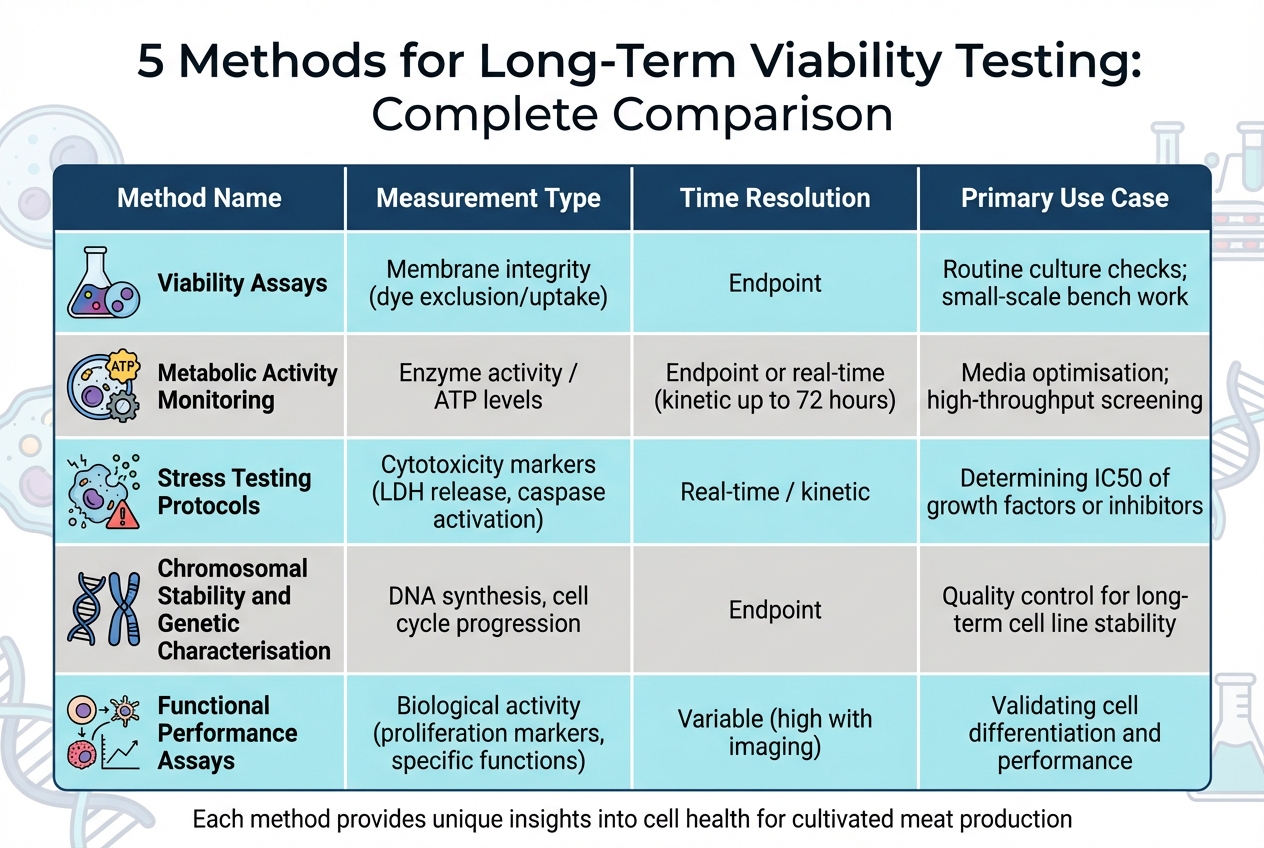
Comparison of 5 Long-Term Viability Testing Methods for Cultivated Meat Production
The table below outlines the main features of five methods used for long-term viability testing, highlighting their measurement types, time resolution, and typical applications.
| Method | Measurement Type | Time Resolution | Primary Use Case |
|---|---|---|---|
| Viability Assays | Membrane integrity (dye exclusion/uptake) | Endpoint | Routine culture checks; small-scale bench work |
| Metabolic Activity Monitoring | Enzyme activity / ATP levels | Endpoint or real-time (kinetic up to 72 hours) | Media optimisation; high-throughput screening |
| Stress Testing Protocols | Cytotoxicity markers (LDH release, caspase activation) | Real-time / kinetic | Determining IC50 of growth factors or inhibitors |
| Chromosomal Stability and Genetic Characterisation | DNA synthesis, cell cycle progression | Endpoint | Quality control for long-term cell line stability |
| Functional Performance Assays | Biological activity (proliferation markers, specific functions) | Variable (high with imaging) | Validating cell differentiation and performance |
This comparison highlights the unique advantages and limitations of each method, particularly in terms of measurement type and timing. Time resolution plays a key role in tracking long-term changes, with non-lytic assays allowing continuous monitoring for up to 72 hours [2].
In 3D culture models - commonly used in cultivated meat production - traditional colourimetric assays often face challenges with reagent penetration. Specialised reagents designed for 3D systems, featuring stronger detergents, are necessary to ensure effective assay performance. Methods that track markers released into the medium, such as LDH in stress testing, are particularly useful for reaching the core of microtissues [2]. Combining viability assays with cytotoxicity testing provides a clearer distinction between cytostatic (growth-inhibiting) and cytotoxic (cell-killing) effects [2][6]. This concise overview helps guide method selection to suit a variety of testing needs.
Conclusion
To ensure accurate and reliable results in testing cell line viability, a multifaceted approach is essential. Long-term viability testing in cultivated meat production requires the use of multiple assays, as each evaluates different cellular markers. Relying on just one parameter can lead to misleading results - a cell might appear viable but could be metabolically inactive or even senescent [2].
As Johanna Lee and Mariel Mohns from Promega Corporation explain:
"Choosing a cell health assay method to suit your needs requires an understanding of what each assay is measuring as a marker, how the measurement correlates with cell viability and what are the limitations." [2]
This is especially important when working with 3D scaffolds, where combining orthogonal methods becomes critical [2]. Multiplexing assays within a single well not only improves statistical reliability but also helps conserve valuable cell types [2]. By using this approach, researchers can differentiate between "live", "dead", and "dying or damaged" cells, ensuring more comprehensive validation of experimental outcomes [3]. Moreover, experimental compounds can sometimes alter cellular metabolism or proliferation without causing cell death. Pairing viability assays with toxicity tests helps avoid misinterpretation of such metabolic changes [6].
For companies in the cultivated meat sector, developing robust testing protocols also hinges on access to specialised equipment. Tools like multi-mode microplate readers and automated cell counters enhance assay flexibility and minimise errors [4][5]. Platforms such as Cellbase simplify this process by connecting R&D teams and production managers with verified suppliers. These suppliers offer advanced tools, including microplate readers with temperature and CO₂ controls for real-time monitoring [4] and reagents specifically designed for 3D culture systems. Adopting these integrated methods strengthens the foundation for scalable and reliable cell line performance in cultivated meat production.
FAQs
How often should long-term viability testing be done during passaging?
The frequency of testing for long-term viability during cell passaging varies depending on the protocol and the specific cell line being used. Viability assays are usually conducted at intervals that align with the culture conditions - this could mean testing before each passage or at predetermined time points. For cultivated meat cell lines, regular viability testing is crucial to ensure the cells remain healthy and functional over extended periods of cultivation.
Which assays are most effective for 3D scaffolds with limited reagent diffusion?
When working with 3D scaffolds where reagent diffusion is limited, the acid phosphatase assay (APH) proves to be a reliable choice. This assay performs effectively with spheroids up to 650 µm and even 900 µm in size, without requiring dissociation.
Additionally, cell viability assays specifically designed for 3D constructs are highly compatible with these conditions. These assays are sensitive enough to account for the diffusion challenges inherent in 3D scaffolds, making them particularly suited for assessing long-term cell viability. This makes them an excellent tool for research in cultivated meat, where maintaining cell health over time is crucial.
What is the minimum set of orthogonal tests to confirm a cell line is safe and stable?
To confirm a cell line's safety and stability, a few essential tests are required. These include genetic stability assessments such as karyotyping, SNP arrays, or next-generation sequencing. These methods help identify mutations or chromosomal abnormalities that could compromise the cell line.
Additionally, contamination testing and cell authentication play a crucial role. These tests ensure the cell line's identity and purity, reducing the risk of cross-contamination or misidentification. When combined, these procedures safeguard both the genetic integrity and overall safety of cell lines used in cultivated meat production.


